Platform
MicroCell
Optical Microscopy and Flow Cytometry Core Facility
Presentation
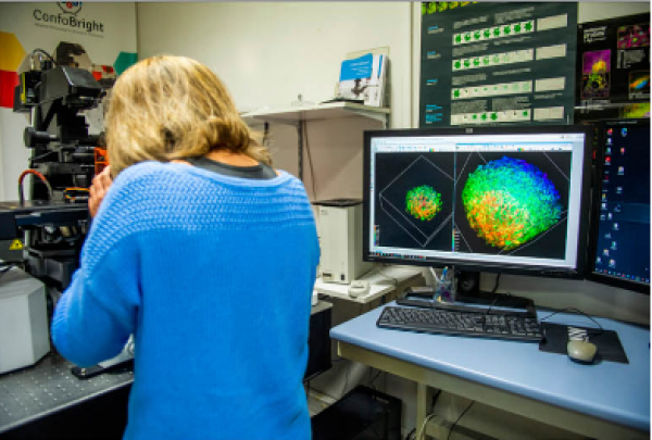
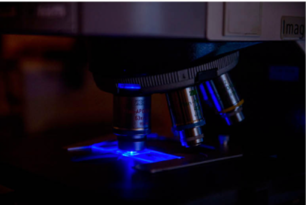
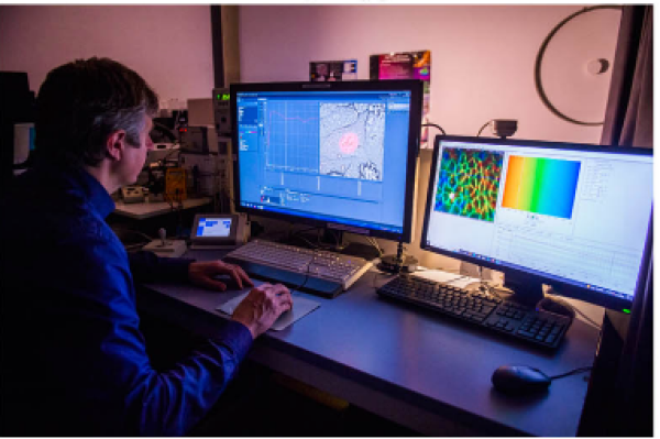
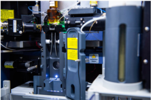
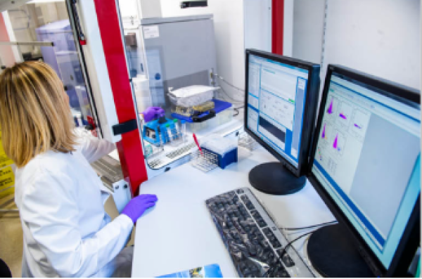
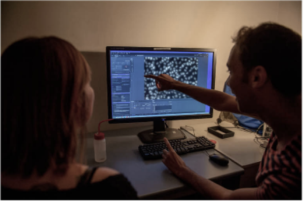
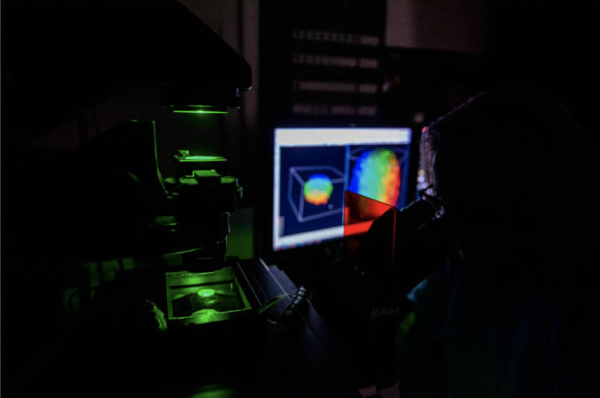
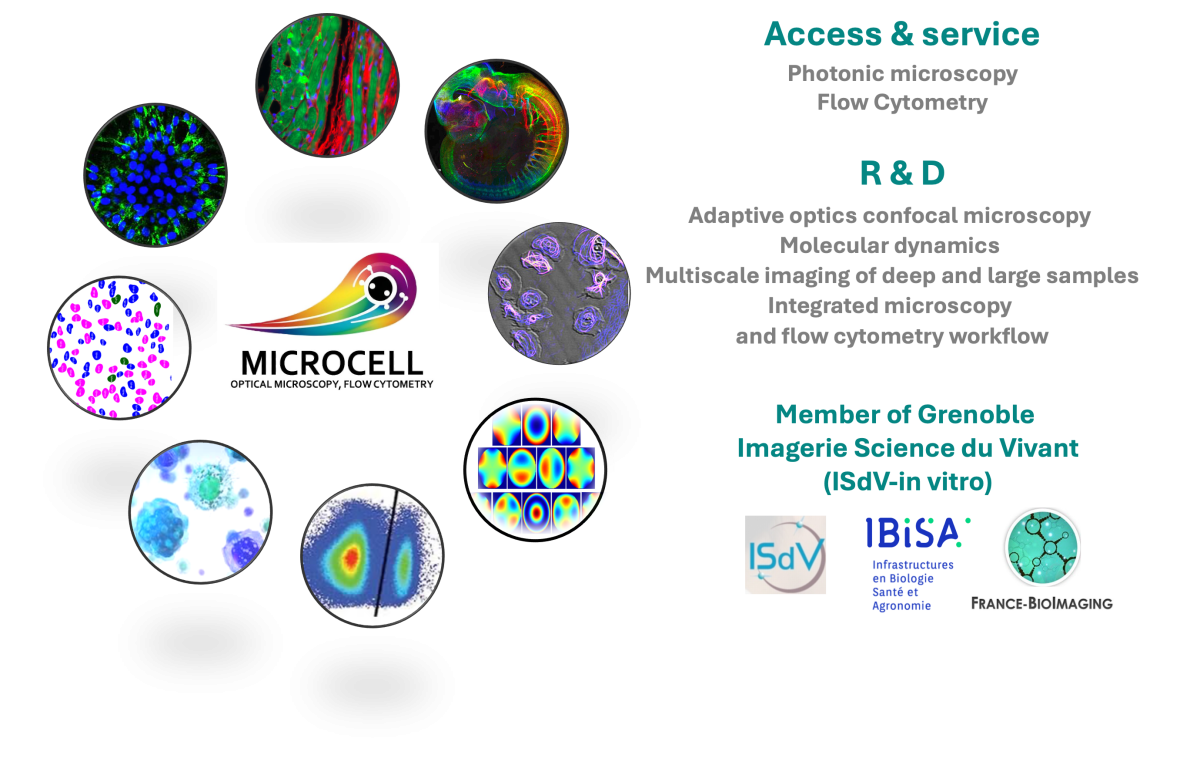
The Optical Microscopy and Flow Cytometry Platform (MicroCell) offers a wide range of research instruments, technical training and technological innovations in cellular imaging. Since 2009, it has been recognized by the national GIS IBiSA label and it is part of the Grenoble multisite platform Live Sciences Imaging - In vitro (ISdV). The high-end instruments and staff expertise are made available to IAB research teams and also accessible to the entire scientific community for public research laboratories or private companies via Floralis, the university management subsidiary. The main specificity of this core facility is the integration, combination and validation of complementary optical microscopy techniques and flow cytometry modalities in the frame of methodological developments in biology.
A steering committee supports the platform to define trans-team scientific and technological development plans, to represent the platform in the decision-making committees of the IAB and its partners and also optimally position the platform in local, regional and international strategies.
Steering Committee composition : Olivier Destaing, DR-CNRS, Zuzana Macek-Jilkova, IR-CHU Grenoble Alpes, Amina Touré, DR-CNRS, Antoine Delon, Pr UGA
Centers of expertise
The Optical Microscopy and Flow Cytometry Platform (MicroCell) offers a wide range of research instruments, technical training and technological innovations in cellular and tissue imaging at the micrometric and sub-micrometric scales.
Learn more
Our methodological developments aim to overcome technological barriers and develop new tools and methods in photonic imaging. Seeing beyond what was visible and in a more quantitative and better controlled manner in space and time allows us to improve the understanding of normal and pathological cellular mechanisms.
Learn more
Our platform provides the environment necessary for the implementation of this high-throughput multiparametric method to characterize cellular subpopulations and decipher their relationships. Flow cytometry also makes it possible to study the functional state of cells, cytokine production and to monitor cell death and survival or to detect rare events while offering the possibility of isolating and recovering cells of interest.

