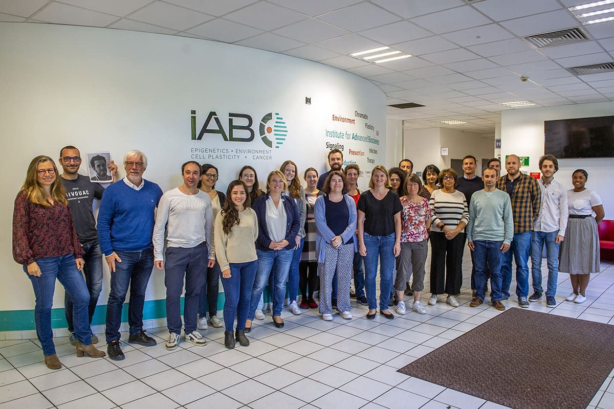
Équipe
Thérapie ciblée, diagnostic précoce et imagerie du cancer
Dpt: Microenvironnement, Plasticité cellulaire et Signalisation
Nos activités de recherche
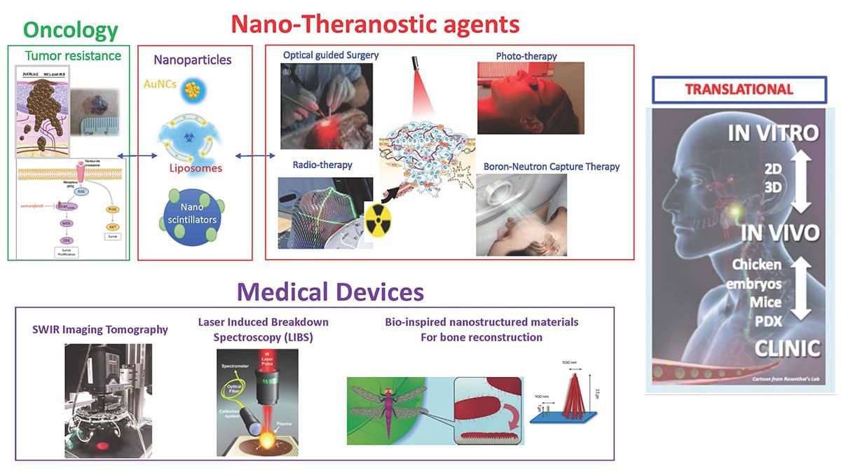
Notre objectif principal est de définir des nano-vecteurs théranostiques pour le traitement du cancer, et en parallèle de développer les dispositifs médicaux adaptés.
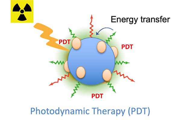
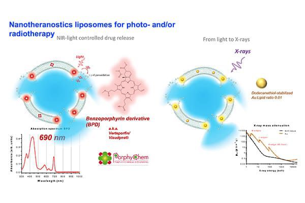
Les nanoparticules (NP) que nous développons sont pour la plupart détectables en imagerie optique proche-infrarouge (NIR I et NIR II (ou SWIR)). Leur activité thérapeutique est, quant à elle, définie pour échapper aux mécanismes de résistances habituellement mis en place par les tumeurs. Ainsi, ces NP sont élaborées pour optimiser l’accumulation des drogues dans les tumeurs et/ou pour potentialiser l’effet de la photo- et thermo-thérapie sous excitation lumineuse ou de la radiothérapie sous excitation par rayons X ou neutrons (thérapie par capture de neutrons).
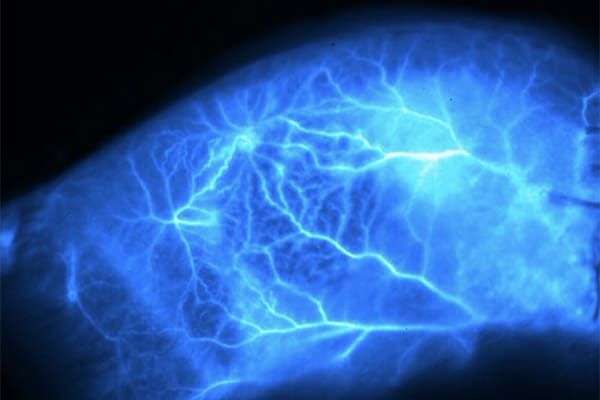
Les NP fluorescentes dans le NIR/SWIR sont également utilisées par notre équipe de chirurgiens oncologues et de chirurgie de reconstruction pour réaliser des opérations de chirurgie guidées par l’optique.
En parallèle, nous développons les technologies pour détecter et suivre ces NP grâce à des instruments de diagnostique (e.g. LIBS ; Laser Induced Breakdown Spectroscopy) ou de visualisation in vivo (e.g. Fluorescence Reflectance Imaging, Tomographie NIR et SWIR).
Nos nano-vecteurs, méthodes et instruments sont évalués in vitro en 2D et 3D (microtumeurs ± microfluidique) puis testés sur des modèles d’embryon de poulet ou en préclinique chez des modèles de rongeurs avant leur transfert éventuel en clinique ou vers l’industrie. L’ensemble de ces nouvelles technologies est aussi mis à la disposition de la communauté scientifique via notre plateforme d’imagerie du petit animal (OPTIMAL) reconnue au niveau national dans le cadre des programmes IBISA et France Life Imaging (FLI).
Notre recherche est effectuée dans un environnement interdisciplinaire où les biologistes travaillent en étroite collaboration avec des chimistes, des physiciens, des sociétés de biotechnologie et des équipes cliniques.
Nos axes de recherche
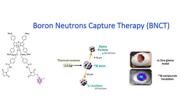
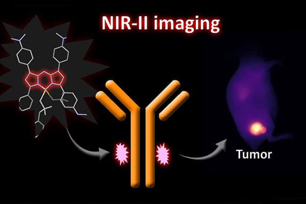
Nous concevons différents types de NP fluorescentes, certaines peuvent être détectées dans les fenêtres NIR I ou NIR II. Les effets anticancéreux de ces nano-vecteurs sont déclenchés par l'excitation par rayons X, par neutrons ou par de la lumière délivrés sur le site tumoral. Ces NP sont soit des nanoclusters métalliques, des nanoscintillateurs, des NP contenant du bore ou des liposomes activables qui peuvent induire une augmentation de la dose de rayons X (RDE), une radiosensibilisation, une thérapie par capture de neutrons par le bore (BNCT)…
En savoir plusNous caractérisons les interactions entre les cellules tumorales et leur microenvironnement afin d’identifier des cibles thérapeutiques et de définir de nouvelles thérapies et/ou combinaisons thérapeutiques. Nous étudions notamment les voies EGFR, IGFR et intégrines et nous développons des activités de recherche fondamentale, appliquée et translationnelle vers la clinique en nous appuyant sur le service de dermatologie du CHU Grenoble Alpes.
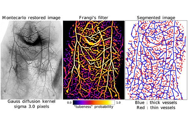
En savoir plusNous développons des instruments d’imagerie dans le proche infrarouge (NIR) pour les applications in vivo. Les fenêtres NIR I et NIR II permettent de sonder les processus physiologiques et moléculaires à haute résolution. Pour le diagnostic, nous développons la microscopie élémentaire LIBS (laser induced breakdown spectroscopy) pour déterminer la composition chimique des tissus (et en particulier détecter nos NP) sur des lames ou biopsies.
En savoir plusNos publications majeures
Voir toutes les publicationsNos activités en images
Nos technologies
- Culture cellulaire 2D et 3D (sphéroïdes et organoïdes)
- Modèle in vivo sur membrane chorioallantoïde (CAM) de poulet
- Modèles animaux
- Imagerie NIR in vivo
- Chirurgie guidée par l’optique
- Radio, photo, neutron thérapies
- Microscopie Multi-élémentaire LIBS sur tissus En savoir plus